![]() Figure 6 of
Cerra, Mol Vis 2003;
9:689-700.
Figure 6 of
Cerra, Mol Vis 2003;
9:689-700.
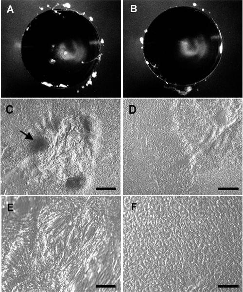
Figure 6. Inhibition of TGFβ-induced changes by PSS
Lenses from immature rats were cultured with 1.2 ng/ml TGFβ (A,C,E) or with TGFβ plus 0.2 μM PSS (B,D,F) for 4 days. Micrographs show a single representative lens from each treatment group. The lenses were photographed at the end of the culture period (A,B) then whole mounts were prepared as described in the legend to Figure 2, fixed, and photographed using Integrated Modulation Contrast. Discrete anterior opacities are visible in the central region of the lens cultured with TGFβ (A) but not in the lens cultured with TGFβ plus PSS (B); the crescent-shaped cloudy area in both lenses is an artifact due to glare from the light source. With TGFβ alone, plaques formed (C, dark areas, arrow) and regions between plaques showed extensive spindle cell formation (E). When PSS was included with TGFβ, plaque formation was suppressed (D) and most of the cells retained normal epithelial morphology (F). Bar represents 500 μm in A and B, 200 μm in C and D, and 50 μm in E and F.
