![]() Figure 2 of
Cerra, Mol Vis 2003;
9:689-700.
Figure 2 of
Cerra, Mol Vis 2003;
9:689-700.
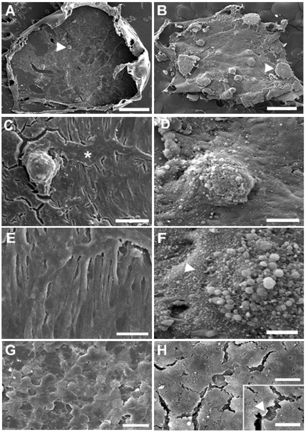
Figure 2. Effect of FGF on plaque formation induced by TGFβ: SEM
Lenses from weanling rats were cultured with 1.5 ng/ml TGFβ in the absence (A,C,E) or presence (B,D,F) of 20 ng/ml FGF. Controls were cultured without addition of growth factors (G) or with FGF alone (H). At the end of the 4 day culture period, the anterior capsule with adhering cells and plaques was peeled off and processed for scanning electron microscopy (SEM). After culturing with TGFβ alone only a few small plaques were present (A, arrowhead), whereas culturing with TGFβ and FGF induced the formation of numerous moderate to very large plaques (B), the larger ones being located in the more peripheral region (arrowhead). With TGFβ alone, plaques were relatively smooth in appearance and rose abruptly from the surrounding cells (C), many of which were elongated into spindle-like forms and in parallel arrays (C,E). Plaques were often associated with "channels" of non-elongated cells traversing regions of spindle cells (C, asterisk; E). When FGF was included with TGFβ, plaques appeared to emerge more gradually out of generally thickened regions of cells (D,F). Cells were generally less attenuated and more closely apposed than with TGFβ alone. In addition, exposed cellular surfaces were often coated with a meshwork of ECM-like material (F; arrowhead) and plaques exhibited rounded protrusions at their apex, giving them a rough appearance (D,F). Epithelial cells in the lens cultured without growth factors (G) retained a loose cobblestone arrangement. After culturing with FGF alone (H), some enlargement of cells was evident with formation of tongue-like processes (inset, arrowhead). Bar represents 500 μm in A and B, 50 μm in C and D, 20 μm in E-H, and 10 μm in the inset of H.