![]() Figure 4 of
Cerra, Mol Vis 2003;
9:689-700.
Figure 4 of
Cerra, Mol Vis 2003;
9:689-700.
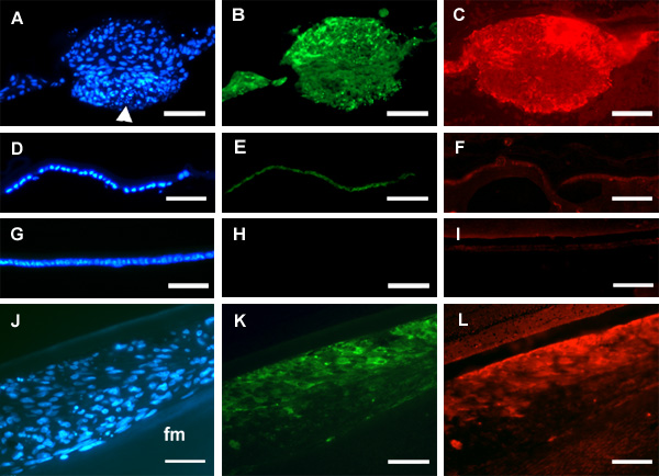
Figure 4. Immunolocalization of myofibroblast and fibroblast markers in lenses cultured with TGFβ and FGF
Lenses from young rats (A-I) and adult rats (J-L) were used. All images are presented lens capsule surface uppermost. A-C: Lenses from weanling rats were cultured for 4 days with 1.5 ng/ml TGFβ and 20 ng/ml FGF, then whole mounts were embedded in paraffin and sectioned, as described in the legend to Figure 3. Hoechst staining (A) revealed a large plaque. Some nuclei were pyknotic especially in the tightly packed apical region (arrowhead). Reactivity for α-smooth muscle actin was detected in this section (B). Type I collagen reactivity was detected in an adjacent section (C). Reactivity was strongest in the subcapsular region of the plaque and adjacent multilayered cells. D-I: Sections of whole mounts from corresponding control lenses cultured without growth factors (D-F) or with 20 ng/ml FGF alone (G-I). Regions corresponding to the Hoechst-stained monolayers depicted in D and G showed negligible reactivity for α-smooth muscle actin (E,H), type I collagen (F) and fibronectin (I). J-L: Lenses from adult female rats were cultured for 8 days with 3 ng/ml TGFβ and 30 ng/ml FGF, fixed intact, embedded in paraffin and sectioned. Hoechst staining revealed a continuous, thickened anterior layer of cells (J). Many cells in this section exhibited reactivity for α-smooth muscle actin (K) and fibronectin (L), which was particularly strong in the region underlying the lens capsule. No reactivity was detectable in the underlying fiber cell mass (fm). Bar represents 60 μm in A-I and 40 μm in J-L.