![]() Figure 3 of
Cerra, Mol Vis 2003;
9:689-700.
Figure 3 of
Cerra, Mol Vis 2003;
9:689-700.
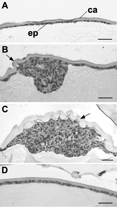
Figure 3. Effect of FGF on plaque formation induced by TGFβ: H&E staining
Lenses from weanling rats were cultured for 4 days with 1.5 ng/ml TGFβ alone (A,B), with TGFβ plus 20 ng/ml FGF (C) or with FGF alone (D). Whole mounts of epithelial regions were prepared, as described in the legend to Figure 2, embedded in paraffin and sectioned. In lenses cultured with TGFβ alone, most of the epithelium retained typical lens epithelial morphology, that is, it consisted of a monolayer of cuboidal cells (A), although patchy multilayering was present in some regions (not shown). B shows the only plaque that formed in the lens depicted in A, located at the anterior pole; some wrinkling of the lens capsule was evident (arrow). When FGF was included with TGFβ, the numerous plaques that formed were much larger (C) and severe wrinkling of the capsule occurred (arrow). Cells were more tightly packed in the region adjacent to the fiber mass. In lenses cultured with FGF alone, the epithelium remained as a monolayer (D). Bar represents 20 μm. The lens capsule (ca) and lens epithelium (ep) are labelled.