![]() Figure 2 of
Cox, Mol Vis 2003;
9:665-672.
Figure 2 of
Cox, Mol Vis 2003;
9:665-672.
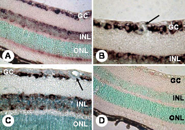
Figure 2. Localization of PDGF-B mRNA expression
Hybridization with PDGF-B riboprobes shows a largely similar localization pattern to that observed with PDGF-A (see Figure 1). Non-diabetic (A, B) and diabetic murine retina (C) shows mRNA expression in the ganglion cell layer (GC), the innermost aspect of the inner nuclear layer (INL) and to a small, extent in the inner segments of the photoreceptors (A). When retinas from non-diabetic and diabetic mice are compared, there are no obvious differences in reaction product localization or intensity (compare B with C) and there is no PDGF-B mRNA in the retinal blood vessels (B, C; arrows). Sense riboprobes showed no localization of mRNAs (D). Original magnifications: x400 (A, D) and x100 (B and C). The outer nuclear layer (ONL) is also labeled in the micrograph.