![]() Figure 1 of
Cox, Mol Vis 2003;
9:665-672.
Figure 1 of
Cox, Mol Vis 2003;
9:665-672.
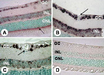
Figure 1. Localization of PDGF-A mRNA expression
In situ hybridization in retinal sections from non-diaebtic (A and B) and diabetic mice (C) with PDGF-A riboprobes shows mRNA expression in the inner retina, specifically localized to the ganglion cell layer (GC) and both the inner and outer aspects of the inner nuclear layer (INL). In the outer retina there is expression localized to the inner segments of the photoreceptor cells (A; arrow). When retinas from non-diabetic and diabetic mice are compared, there are no obvious differences in reaction product localization or intensity (compare B with C). It is notable that the retinal blood vessels (B, C; arrows) show no expression of PDGF-A. Negative hybridization with sense riboprobe is shown in D. Original magnifications: x400 (A, D) and x100 (B, C). The outer nuclear layer (ONL) is also labeled in the micrograph.