![]() Figure 3 of
Liljekvist-Larsson, Mol Vis 2003;
9:657-664.
Figure 3 of
Liljekvist-Larsson, Mol Vis 2003;
9:657-664.
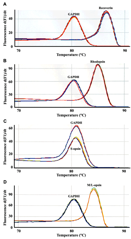
Figure 3. Specificity of amplification reactions
Melt curve analysis of the amplification reactions in the real-time PCR. Melting curves of recoverin (A), rhodopsin (B), S-cone opsin (C) and M/L-cone opsin (D) in cultured and control retinas. Each analysis also shows a melt curve analysis of each target amplified with GAPDH in cultured and control retinas. The peaks in the first derivate plot indicate the presence of a specific melting product for each target and GAPDH.