![]() Figure 2 of
Liljekvist-Larsson, Mol Vis 2003;
9:657-664.
Figure 2 of
Liljekvist-Larsson, Mol Vis 2003;
9:657-664.
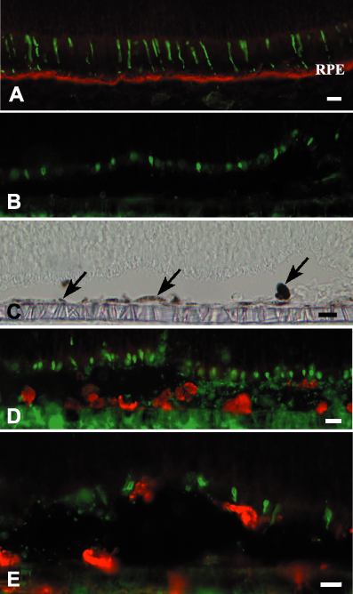
Figure 2. Photoreceptor outer segments and retinal pigment epithelium
(A) PNA-positive (green) cone matrix sheaths are closely associated with the retinal pigment epithelium (RPE; red) in normal P14 retina. The RPE is immunolabeled for RPE65. (B) In cultured retinas, the cone matrix sheaths are rudimentary but distinctly PNA-positive. Note the absence of immunolabeling for RPE65 in the RPE. (C) Light microscopy of the same field as shown in B, showing the presence of several RPE cells with distinct pigmentation (arrows). (D) Labeling with anti-ED1 (red) shows the presence of activated macrophage-like cells in the RPE-layer. Cone matrix is labeled for PNA (green). (E) High magnification of PNA-labeled (green) cone matrix sheaths approached by ED1-positive (red) activated macrophage-like cells. Scale bars represent 15 μm in A-D and 10 μm in E.