![]() Figure 1 of
Liljekvist-Larsson, Mol Vis 2003;
9:657-664.
Figure 1 of
Liljekvist-Larsson, Mol Vis 2003;
9:657-664.
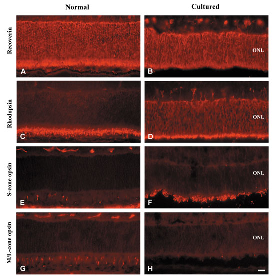
Figure 1. Immunolabeling of in vivo and cultured retinas
Immunofluorescent labeling of photoreceptor cells and outer segments in normal P14 retina and cultured retinas. Photoreceptor cells in P14 (A) and 11 days in vitro (B) retina are immunolabeled for recoverin. Note that the outer nuclear layer (ONL) of P14 retina is considerably thicker. Immunolabeling for rhodopsin accumulates in photoreceptor outer segments at P14 (C). In cultured retina, rhodopsin expression is present in the outer segments and intense labeling is also evident in the entire ONL and in cell somata located at the vitreal aspect of this layer (D). Compared to the P14 retina (E), numerous cones are immunolabeled for S-cone opsin in the 11 days in vitro retina (F). The density of cone photoreceptor cells immunolabeled for M/L-opsin is much higher in the P14 (G) than in the 11 days in vitro retina (H). Scale bar represents 15 μm.