![]() Figure 5 of
Srivastava, Mol Vis 2003;
9:644-656.
Figure 5 of
Srivastava, Mol Vis 2003;
9:644-656.
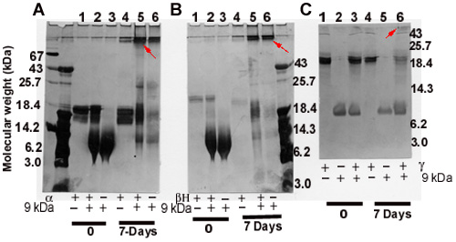
Figure 5. SDS-PAGE analysis of crosslinked species produced following incubation of the 9 kDa polypeptide with crystallins
A: α-Crystallin (15 μg) and 9 kDa polypeptide (15 μg). B: βH-crystallin (6 μg) and 9 kDa polypeptide (15 μg) and C: γ-Crystallin (10 μg) and 9 kDa polypeptide (10 μg). The constituents of each of the incubated mixture are shown at the bottom of the gel. In these experiments, as apparent from the above quantities, the individual crystallins and the polypeptide were incubated at the ratio of 1:1 or 1:2. The protein species on gels were stained with silver stain.