![]() Figure 3 of
Srivastava, Mol Vis 2003;
9:644-656.
Figure 3 of
Srivastava, Mol Vis 2003;
9:644-656.
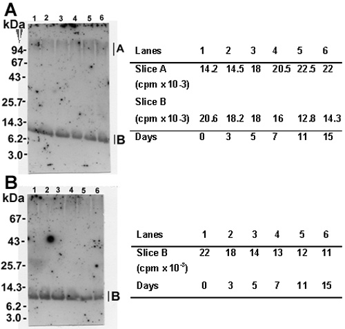
Figure 3. Analysis of crosslinked 9 kDa polypeptide species
Immunoblots following reactivity with: A anti-γ-crystallin antibody and B anti-9 kDa polypeptide antibody. Both blots were reacted with radioiodinated protein A as a secondary antibody to visualize and quantify the immunoreactive proteins. The A and B portions of immunoblot A, and the B portion of immunoblot B were excised and counted for radioactivity. These counts are shown in the adjacent tables. Preparations used for western blot analysis in lanes 1 to 6 included the following samples incubated for the length of time as shown in Figure 2B: lane 1 is day 3, lane 2 is day 7, lane 3 is day 9, lane 4 is day 13, lane 5 is day 15, and lane 6 is day 19.