![]() Figure 5 of
Wride, Mol Vis 2003;
9:360-396.
Figure 5 of
Wride, Mol Vis 2003;
9:360-396.
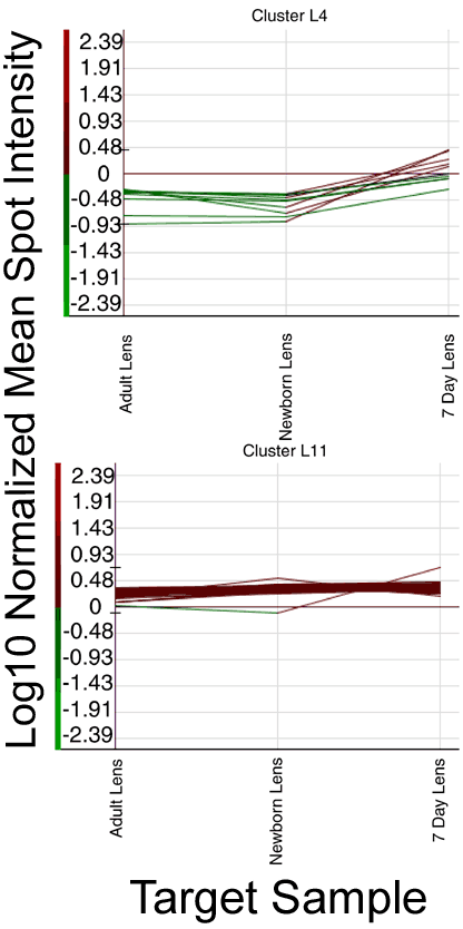
Figure 5. Clusters of genes selected from K-means clustering of the lens samples
Clusters of genes selected from the K-means clustering in Figure 4 potentially differentially expressed in the lens samples (See Table 5 for lists of genes). Y axis shows the log10 of the normalized intensity and the X axis shows the sample. N.B. -0.297 is the mean background intensity for all arrays (only the gene with highest expression at 7 days in cluster L11 was followed up with semi-quantitative RT-PCR).