![]() Figure 6 of
Zhang, Mol Vis 2003;
9:238-248.
Figure 6 of
Zhang, Mol Vis 2003;
9:238-248.
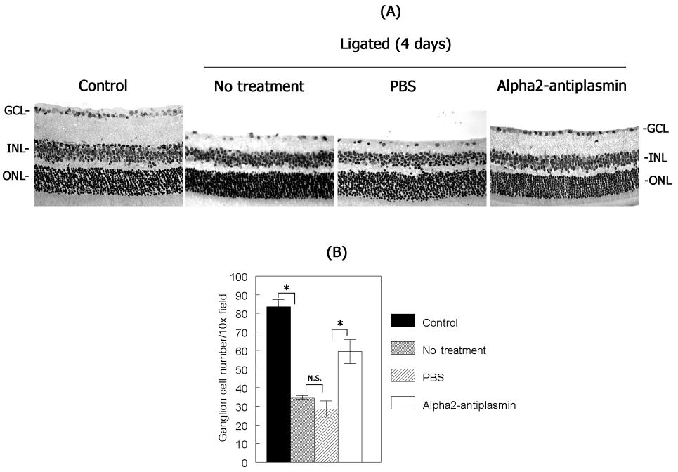
Figure 6. α2-antiplasmin attenuates ganglion cell loss
A. Plasmin inhibitor, α2-antiplasmin was intravitreally injected 10 min before optic nerve ligation and retinal morphology in H & E stained cross sections was assessed 4 days after optic nerve ligation. B. Retinal ganglion cell loss was quantified by counting the cells in the ganglion cell layer in a 10x field at a distance of 1.0-2.0 mm from the optic disc. Data from 8-10 sections from 4 different eyes were analyzed by a paired Student's t-test using Slidewrite software. The bars and error bars represent the mean and standar error of the mean, respectively. The asterisks ("*") indicate a statistically significant difference (p<0.005).