![]() Figure 2 of
Zhang, Mol Vis 2003;
9:238-248.
Figure 2 of
Zhang, Mol Vis 2003;
9:238-248.
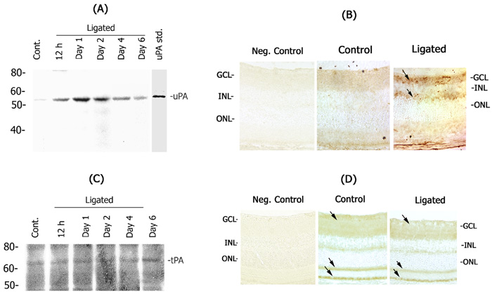
Figure 2. Detection of uPA and tPA in the retina
Retinal extracts were prepared from optic nerve ligated and uninjured control eyes over a time course from 12 h to 1 week after optic nerve ligation. Aliquots containing equal amount of protein (25 μg) were electrophoresed on 10% SDS-polyacrylamide gels, transferred onto PVDF membranes and analyzed by western blotting. Membranes were probed with antibodies against uPA (A) or tPA (C). Immunostaining was performed on 10 μm retinal cross sections using antibodies against uPA (B) and tPA (D). Immunostaining was visualized using appropriate secondary antibodies and an ABC kit. Arrows indicate positive immunostaining. GCL=ganglion cell layer, INL=inner nuclear layer, ONL=outer nuclear layer. (40x magnification)