![]() Figure 1 of
Zhang, Mol Vis 2003;
9:238-248.
Figure 1 of
Zhang, Mol Vis 2003;
9:238-248.
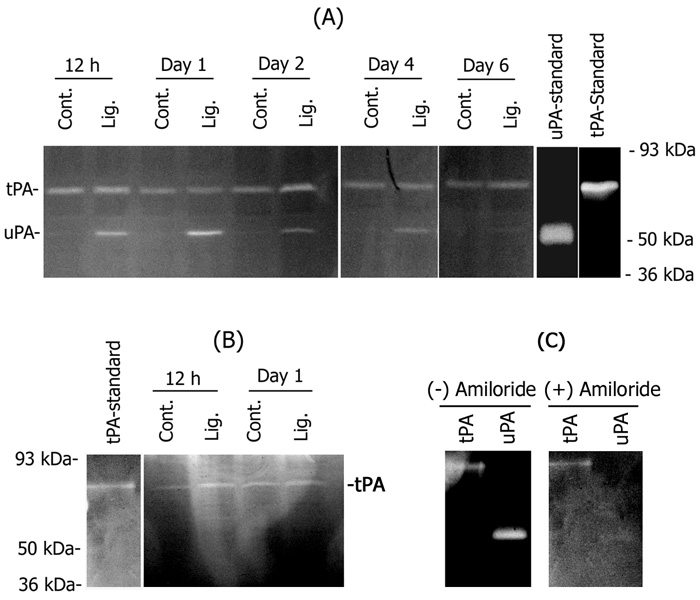
Figure 1. uPA induction in the retina
A. Retinal extracts were prepared from optic nerve-ligated (Lig.) or uninjured control (Cont.) eyes over a time course from 12 h to 6 days after optic nerve ligation (n=4 for each experiment). Aliquots containing equal amount of protein (25 μg) were analyzed by plasminogen/fibrinogen zymography. For comparison, the samples were co-electrophoresed with a sample of purified murine tPA (0.5 ng), murine uPA (0.5 ng), and with reduced molecular weight size standards (not shown). The migration position of the clear bands representing tPA (65 kda) and uPA (55 kDa) are indicated. B. To differentiate between uPA and tPA, equal amount of protein from control and injured retinas extracted 12 h and day 1 after optic nerve ligation were subjected to plasminogen/fibrinogen zymography and the gels were incubated with uPA inhibitor amiloride (200 μM) during activation. For comparison, the samples were co-electrophoresed with murine tPA (0.25 ng). C. To determine the specificity of amiloride to inhibit uPA, known amounts of uPA (1.25 ng) and tPA (0.125 ng) were subjected to plasminogen/fibrinogen zymography and the gels were incubated with or without 200 μM amiloride during activation. Note the inhibition of uPA in the presence of amiloride.