![]() Figure 3 of
Zhang, Mol Vis 2003;
9:231-237.
Figure 3 of
Zhang, Mol Vis 2003;
9:231-237.
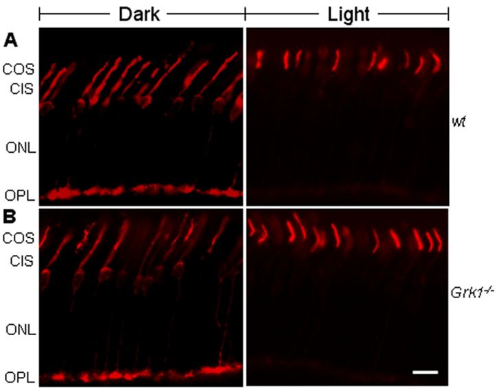
Figure 3. The distribution of cone arrestin in retinas
In dark-adapted (left panels) states, cone arrestin is present in cone outer segments (COS), cone inner segments (CIS), and the outer plexiform layer (OPL) of wild type and Grk1-/- retinas. In the light-adapted (right panels) states, cone arrestin is predominantly in COS. Note that cone arrestin is undetectable in the OPL region (contrast to rod arrestin distribution in Figure 1). A: Wild type (wt) mice. B: Grk1-/- mice. ONL, outer nuclear layer. Bar represents 30 μm.