![]() Figure 1 of
Zhang, Mol Vis 2003;
9:231-237.
Figure 1 of
Zhang, Mol Vis 2003;
9:231-237.
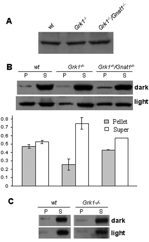
Figure 1. Light-dependent membrane association of rod/cone arrestins in dark-and light-adapted retinas from various genetic backgrounds
A: Comparable levels of rod arrestin are present in wild type (wt), GRK1 knockout (Grk1-/-), and GRK1/Gnat1 (Grk1-/-/Gnat1-/-) double knockout retinas. B: Relative levels of rod arrestin in pellet (P) and in soluble (S) fractions in dark-adapted (upper panel) and light-adapted (lower panel) retinas derived from the indicated genetic backgrounds. One representative set of immunoblots are shown. The amount of arrestin in light-adapted retinas was quantified as described in Methods and shown (bargraph, n=3, error bars represent the standard error of the mean). C: Immunoblots showing the relative amount of cone arrestin in pellet and in soluble fractions from indicated genetic backgrounds. Shown here is one representative experiment from five separate trials. Only small amounts of cone arrestin are present in membrane fractions (P), nearly all cone arrestin is present in the supernatant (S), independent of dark- and light-adaptation or genetic backgrounds.