![]() Figure 5 of
Piriev, Mol Vis 2003;
9:80-86.
Figure 5 of
Piriev, Mol Vis 2003;
9:80-86.
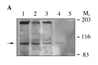
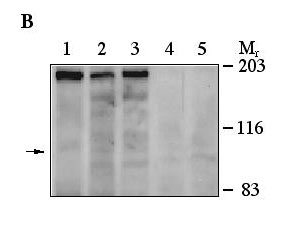
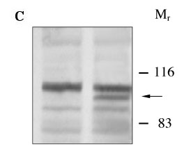
Figure 5. PDEα' expression in HEK293 and Y79 cells
Immunoblots containing protein from bovine cone PDEα' enriched fraction (60 μg total protein, lane 1), Weri-Rb cells (110 μg total protein, lane 2), Y-79 cells (110 μg total protein, lane 3), pNC57-stably transfected HEK293 cells (100 μg total protein, lane 4), and pCIS2-transfected HEK293 cells (100 μg total protein, lane 5). A: Immunoblot incubated with BC18/50 polyclonal peptide antiserum (1:1000 dilution). B: Immunoblot incubated with BC18/50 antiserum and competed with the BC18/50 peptide (10 μg/ml). The arrow indicates the position of the immunoreactive polypeptide identified as the cone-photoreceptor PDE α' subunit. C: Immunoblot probed with antibodies recognizing FLAG peptide. Proteins from non-transfected and transfected Y79 cells are represented in lanes 1 and 2, respectively. The arrow points to the band corresponding to the FLAG-PDEα'. Molecular weight markers are the same as in Figure 4.