![]() Figure 4 of
Piriev, Mol Vis 2003;
9:80-86.
Figure 4 of
Piriev, Mol Vis 2003;
9:80-86.
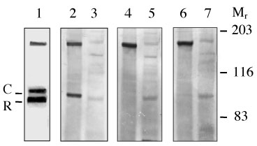
Figure 4. Western blot analysis of the proteins from pC57-transfected CHO cells
Immunoblots containing proteins from a cone PDEα' enriched fraction (60 μg total protein, lanes 1, 2, 4, and 6) and pC57-transfected CHO cells (80 μg total protein, lanes 3, 5, and 7). Lane 1 was incubated with αT1 peptide antiserum (1:1000 dilution) which recognizes rod α and β (R) and cone (C) cGMP-PDE catalytic subunits. Lanes 2 and 3 were incubated with BC18/50 polyclonal peptide antiserum (1:1000 dilution). Lanes 4 and 5 were incubated with BC18/50 antiserum and the BC 18/50 peptide against which the antibody was raised (10 μg/ml). Lanes 6 and 7 were incubated with pre-immune serum from normal rabbit (1:500 dilution). BioRad Laboratories pre-stained molecular weight markers, broad range, were used as standards (Mr): 203 kDa, myosin; 116 kDa, β-galactosidase; and 83 kDa, bovine serum albumin.