![]() Figure 6 of
Zhu, Mol Vis 2002;
8:462-471.
Figure 6 of
Zhu, Mol Vis 2002;
8:462-471.
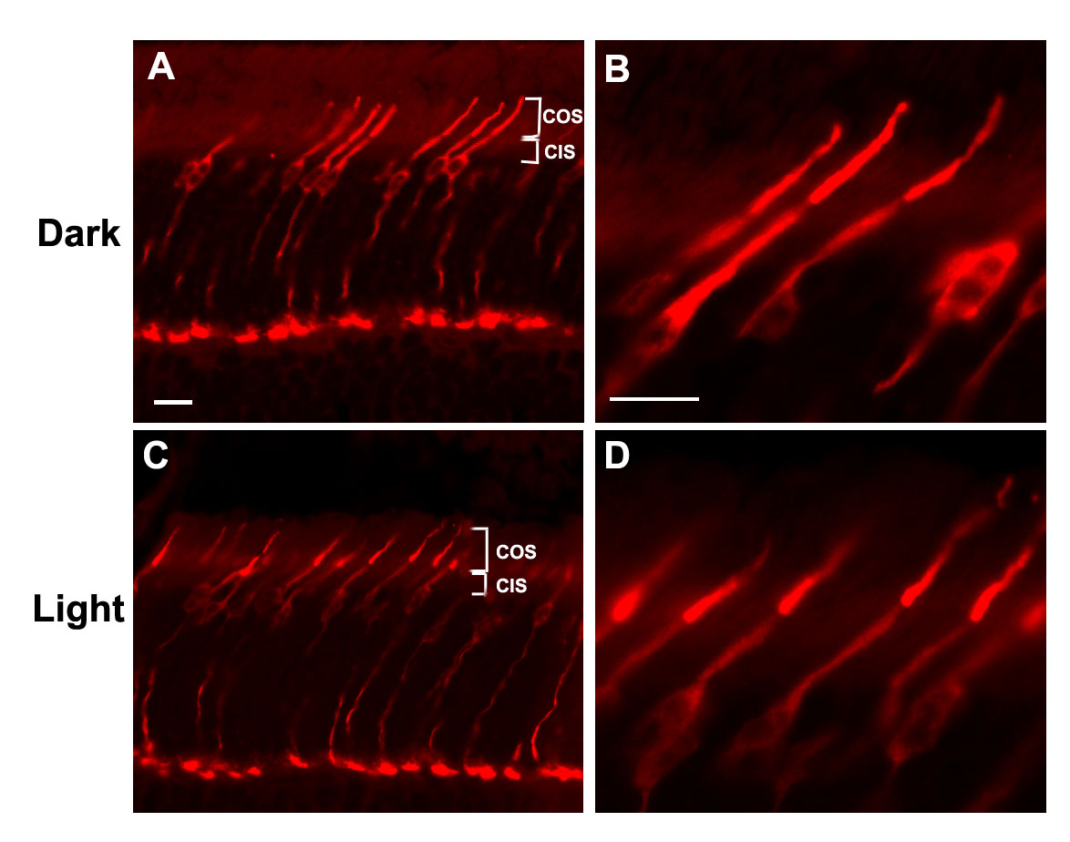
Figure 6. Immunofluorescent staining of adult mouse retinal sections with LUMIJ
Adult C57Bl/6J mice were dark-adapted overnight (dark, A and B) and exposed to light for 4 h (light, C and D) before being killed, and the eyecups were immediately dissected and fixed in 4% paraformaldehyde. Frozen sections were treated as in Figure 5 and stained with the anti-mouse cone arrestin polyclonal antibody LUMIJ (1:1,000) and with Texas Red-conjugated anti-rabbit IgG (1:100). COS, cone outer segments; CIS, cone inner segments. Bar represents 20 μm. Note that mCAR immunoreactivity is evenly distributed throughout the whole cell body in the dark-adapted mouse retina (A and B) but is more intense in the COS in the light-adapted mouse retina (C and D).