![]() Figure 5 of
Zhu, Mol Vis 2002;
8:462-471.
Figure 5 of
Zhu, Mol Vis 2002;
8:462-471.
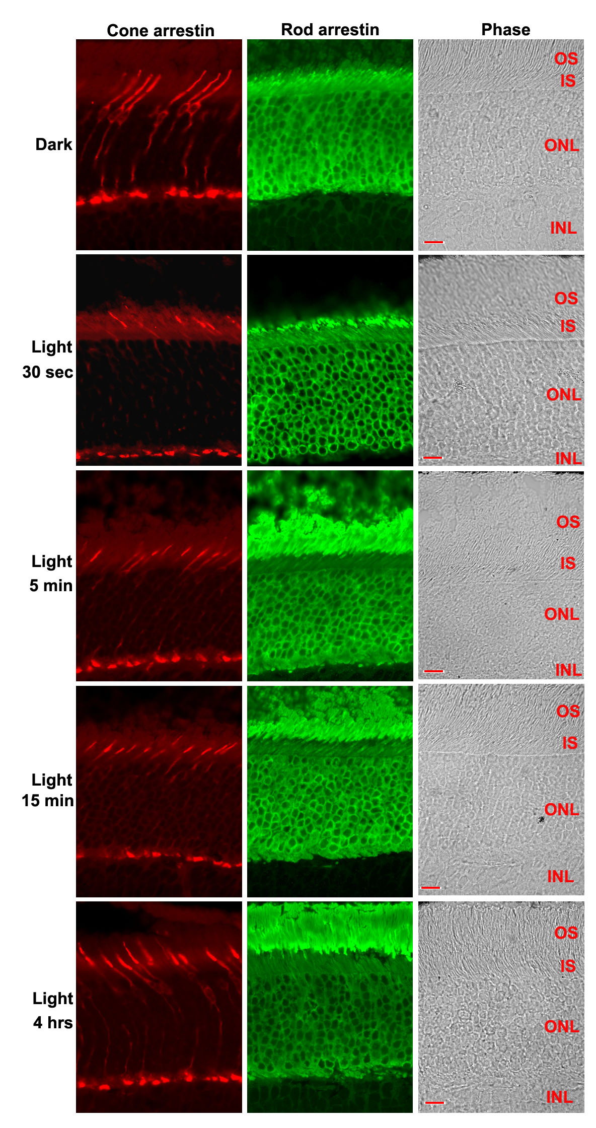
Figure 5. Immunofluorescent staining of adult mouse retinal sections with LUMIJ and C10C10
Adult C57Bl/6J mice were dark-adapted overnight (dark) and exposed to light for selected times before being killed, and the eyecups were immediately dissected and immersion-fixed in 4% paraformaldehyde. Frozen eyes were sectioned at 7 μm through the optic nerve. The sections were heated for 20 min in pre-heated 0.01% sodium citrate buffer (pH 6.0). After blocking, the sections were incubated with the anti-mouse cone arrestin polyclonal antibody LUMIJ (1:1,000) and the anti-rod arrestin monoclonal antibody C10C10 (1:10,000) and then with Texas Red-conjugated anti-rabbit IgG (1:100) and Fluorescin-conjugated anti-mouse IgG. After washing, the sections were examined and photographed as described in methods. OS, outer segments; IS, inner segments; ONL, outer nuclear layer; INL, inner nuclear layer. Bar represents 20 μm.