![]() Figure 5 of
Grossniklaus, Mol Vis 2002;
8:119-126.
Figure 5 of
Grossniklaus, Mol Vis 2002;
8:119-126.
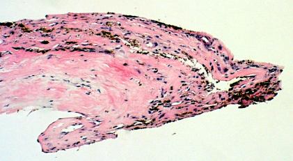
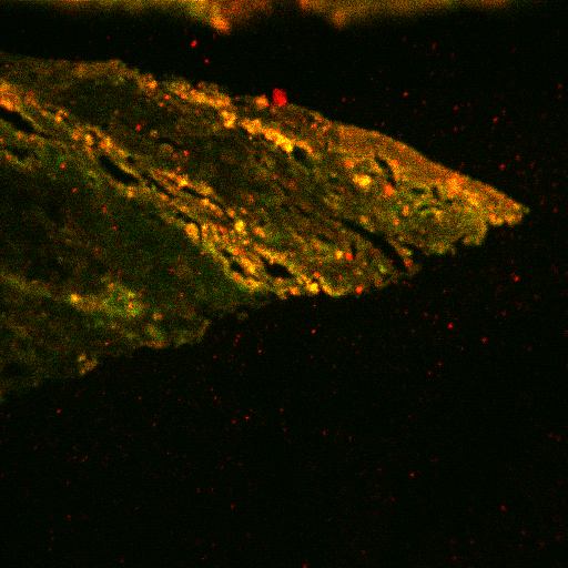
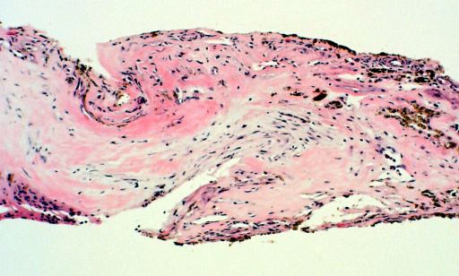
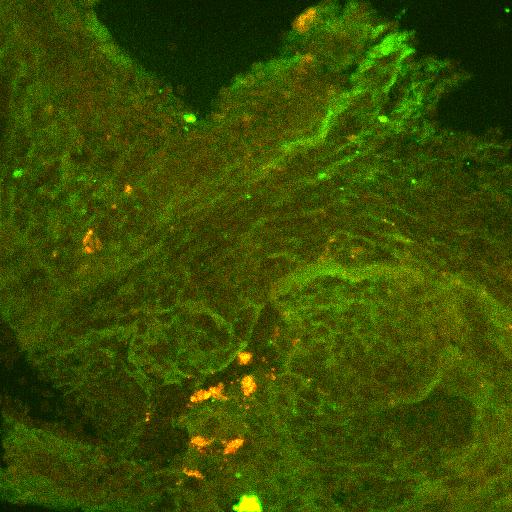
Figure 5. Dual labeling of surgically-excised inflammatory inactive CNV for CK 18 and MC
A: Inflammatory inactive CNV with pigmented cells at edge and fibrosis in stroma. (hematoxylin and eosin, 25x) B: Portion of CNV corresponding to that in Figure 4A showing dual labeling for CK18 and MCP corresponding to pigmented cells at edge, indicating RPE expression of MCP. (FITC, Texas red, 25x) C: Central area of CNV showing more fibrosis than inflammation in stroma. (hematoxylin and eosin, 25x) D: Area of CNV corresponding to Figure 4C showing dual labeling for CD68 and TF in rare stromal cells, indicating rare macrophages expressing TF. (FITC, TF, 63x) E: Negative control. (FITC, Texas red, 63x)
A
B
C
D
E