![]() Figure 4 of
Grossniklaus, Mol Vis 2002;
8:119-126.
Figure 4 of
Grossniklaus, Mol Vis 2002;
8:119-126.
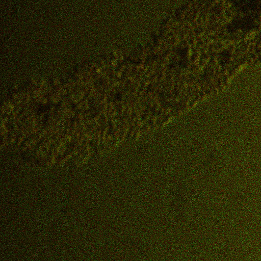
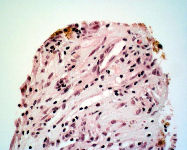
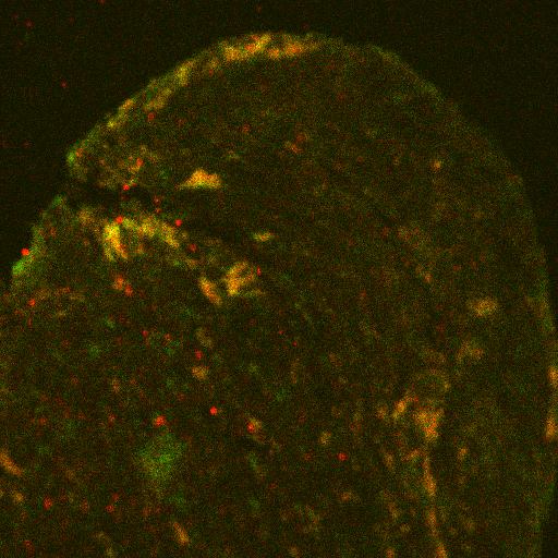
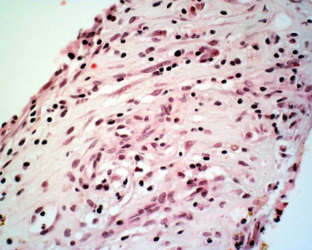
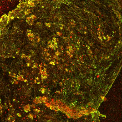
Figure 4. Dual labeling of surgically-excised inflammatory active CNV for CD68 and TF
A: Inflammatory active CNV with pigmented cells (RPE) at edge and inflammatory cells in the stroma. (hematoxylin and eosin, 63x) B: Portion of CNV corresponding to that shown in Figure 3A showing dual labeling for CK18 and MCP along edge, indicating RPE expression of MCP. (FITC, Texas red, 63x) C: Central area of CNV showing numerous inflammatory cells in the stroma. (hematoxylin and eosin, 63x) D: Portion of CNV corresponding to that in Figure 3C showing dual labeling for CD68 and TF, indicating macrophage expression of TF. There are some endothelial cells that express TF. (FITC, Texas red, 63x) E: Negative control (FITC, Texas red, 63x)
A
B
C
D
E