![]() Figure 3 of
Yu, Mol Vis 2001;
7:48-56.
Figure 3 of
Yu, Mol Vis 2001;
7:48-56.
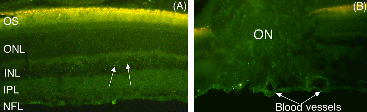
Figure 3. Epifluorescence micrographs of different regions of retinal sections from two eyes 1 h after intravitreal injection of fl-Hsc/Hsp70
A: Fl-Hsc/Hsp70 is apparent in all layers of the retina, but appears to be more prominent in neuropil layers, NFL, IPL, and OPL. In the INL, the arrows indicate vesicular accumulations of the protein. The OS is autofluorescent (compare to Figure 1A), so it was not possible to determine if fl-Hsc/Hsp70 was present there. B: In a section from another eye showing the intraretinal part of the optic nerve, fl-Hsc/Hsp70 can be seen in the walls of two blood vessels, as well as in the neuropil of the optic nerve. 50x magnification. n=13.