![]() Figure 1 of
Yu, Mol Vis 2001;
7:48-56.
Figure 1 of
Yu, Mol Vis 2001;
7:48-56.
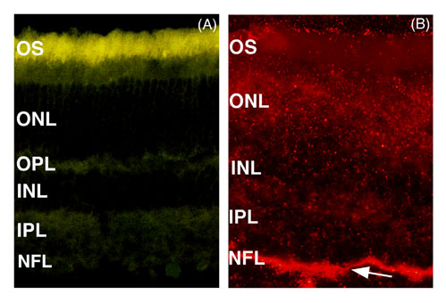
Figure 1. Epifluorescence micrographs of rat retinal sections from control and rhodamine bead-injected eyes
A: Untreated, normal retinal section showing autofluorescence, which appeared as yellow to greenish yellow and was especially bright in the OS. B: Retinal section from an eye taken six h after an intravitreal injection of 10 mg of rhodamine beads. The arrow indicates the high uptake of fluorescent beads in the NFL. Lower levels of diffuse fluorescence can be seen throughout the other retinal layers. In many places, clusters of beads can be seen as punctate fluorescence. 100x magnification. n=4.