![]() Figure 3 of
Pearce-Kelling, Mol Vis 2001;
7:42-47.
Figure 3 of
Pearce-Kelling, Mol Vis 2001;
7:42-47.
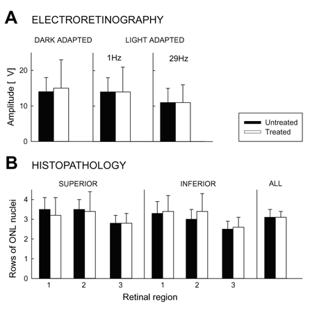
Figure 3. ERG and retinal morphometric comparisons of treated and untreated dogs
ERG and morphometric comparisons of D-cis-diltiazem-treated (white bars) and untreated (black bars) rcd1-affected dogs (14 weeks of age). A: Histograms of average ERG amplitudes for the three measurable responses. B: Histograms of average number of rows of photoreceptor nuclei remaining in rcd1 dogs with or without D-cis-diltiazem treatment. Central (area 1), equatorial (area 2, as shown in Figure 2), and peripheral (area 3) regions were analyzed in both superior and inferior retina. Summary histograms of all data are at right. Error bars represent SD.