![]() Figure 2 of
Pearce-Kelling, Mol Vis 2001;
7:42-47.
Figure 2 of
Pearce-Kelling, Mol Vis 2001;
7:42-47.
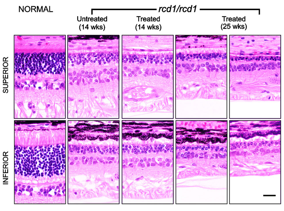
Figure 2. Normal and affected retinal histology
Retinal histology in a representative normal dog (age 21 weeks) and rcd1-affected dogs: one untreated animal (age 14 weeks) and two D-cis-diltiazem-treated animals (ages 14 and 25 weeks). Upper and lower panels represent superior and inferior regions of the retina (area 2), respectively. Calibration bar at lower right, 20 mm.
