![]() Figure 6 of
Chen, Mol Vis 2001;
7:305-313.
Figure 6 of
Chen, Mol Vis 2001;
7:305-313.
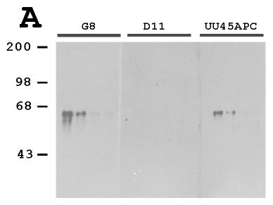
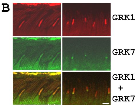
Figure 6. Coexistence of GRK1 and GRK7 in human cone outer segments
A: Western Blot analyses of two fold serial dilutions of H5 cell extracts (10, 5, 2.5, 1.25 mg from left to right) containing recombinant hGRK7 using various antibodies as indicated. The monoclonal anti-GRK1 antibody G8 (1:5,000 dilution) recognized GRK7 while GRK1 specific antibody D11 (at 1:1,000 dilution) did not. UU45APC was used at 1:3,000 dilution. B: fluorescent double labeling of human retinas using D11 (1:500 dilution) and UU45APC (1:1,000 dilution) on sections of an adult retina described in Figure 4C-E (left panels) and of a 4 month old retina described in Figure 4F-H (right panels). Rhodamine conjugated anti-mouse antibody was used to localize D11 and FITC conjugated anti-rabbit antibody was used to localize UU45APC. Upper panels: GRK1 signal (red), middle panels: GRK7 signal (green) and lower panels: co-localization of GRK1 and GRK7 in the cone outer segments (yellow). Images were taken and analyzed using a Zeiss LSM510 confocal microscope.