![]() Figure 2 of
Riley, Mol Vis 2001;
7:297-304.
Figure 2 of
Riley, Mol Vis 2001;
7:297-304.
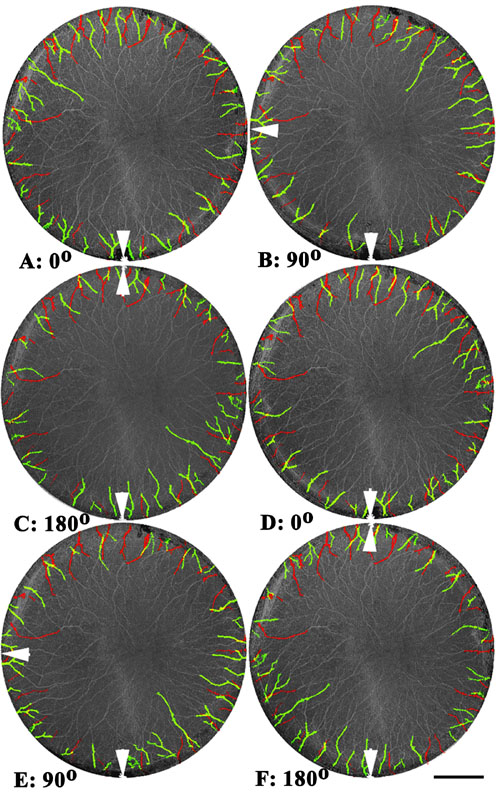
Figure 2. Overlays of mirror images and rotated images of corneal nerves
E16 Japanese quail corneas (6 corneas from 3 embryos) were marked so that right and left corneas from the same embryo could be identified. Reflection test: Images of the nerves were recorded as in Figure 1 and overlaid in register by the choroid fissure. Images of right and left corneas from the same embryo were overlaid at exact positions according to the ventral pole marker (choroid fissure, arrow) to check for innervation specificity (A). Where nerve positions are specific, a third pseudo-color appears (yellow). 90° and 180° rotations (B and C) of one cornea with respect to the other were performed to check if the number of yellow, color-changed nerves diminished. Each rotation showed approximately the same number of color-changed innervation sites, suggesting that innervation sites are not specific, but occur at approximately equal spacing around the circumference of the cornea. Since right and left corneas can be viewed as mirror images, one cornea was flipped around the dorsal-ventral axis and overlaid onto the other corresponding cornea (whose image was not flipped, D); 90° and 180° rotation tests then were performed (E and F). The frequency of color-changed (yellow) nerves stays approximately the same in comparisons of non-rotated (D) and rotated (E and F) images, suggesting, as in Figure 1, that corneal innervation does not occur at specific locations, but rather at uniformly spaced radii around the entire circumference of the cornea. Bar represents 500 mm.