![]() Figure 4 of
Boulanger, Mol Vis 2001;
7:283-287.
Figure 4 of
Boulanger, Mol Vis 2001;
7:283-287.
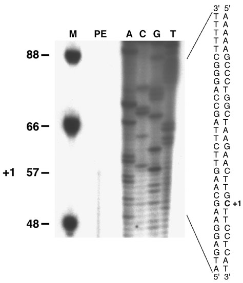
Figure 4. Mapping of the mouse RPE65 mRNA transcription initiation site
The mouse RPE65 mRNA transcription initiation site was mapped by primer extension analysis. RPE/choroid RNA of mice eyes was annealed to the antisense oligonucleotide PE. The extension reaction was also performed without RNA, as a negative control (not shown). The extension product (PE) was analyzed on a 6% denaturing sequencing gel. +1 corresponds to the transcriptional initiation site at the cytosine residue (bold) of the mouse RPE65 gene. The precise position of the 5' end was determined by electrophoresis of the sequence ladder (A, C, T, G) derived from the same antisense primer (T7 Sequenase version 2.1, DNA sequencing kit, Amersham Life Science). The complementary sense strand is shown for clarity. The lane marked M contains the dephosphorylated fX174 Hinf I markers.