![]() Figure 2 of
Boulanger, Mol Vis 2001;
7:283-287.
Figure 2 of
Boulanger, Mol Vis 2001;
7:283-287.
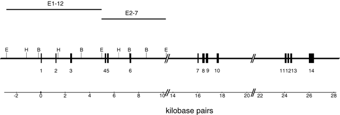
Figure 2. Organization of the mouse Rpe65 gene
A partial restriction map of the 129/Sv mouse Rpe65 gene was derived from sequencing of 2 contiguous subclones (E1-12 and E2-8) of the original P1 clone. Hind III, BamH I and EcoR I sites are shown. No such sites were detected in exons 7-14 or in the sequenced introns beyond exon 7. The large introns between exons 10 and 11 and 13 and 14 were not sequenced. Only the 5' half of the 8 kb intron between exons 6 and 7 was sequenced. The position of exons 1-14 are indicated by the solid boxes, numbered below, with the intervening introns designated by capital letters beneath this. Sequences of exons 7-14 were derived from direct sequencing of PCR products amplified from this P1 clone.
