![]() Figure 4 of
Timmers, Mol Vis 2001;
7:131-137.
Figure 4 of
Timmers, Mol Vis 2001;
7:131-137.
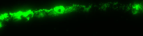
Figure 4. Phagocytotic activity of RPE cells after injection of fluorescent microspheres
Rats were injected with 2 ml of 1 mm fluorescent microspheres and processed as described in Figure 3. Only the fluorescent image is shown. The hexagonal outline typical of RPE cells is visible due to the fluorescent spheres within the cell. In some RPE cells, the nucleus can be seen as the dark spot within the cell. The RPE cells retained their ability for phagocytosis after subretinal injection and consequent retinal detachment.