![]() Figure 3 of
Timmers, Mol Vis 2001;
7:131-137.
Figure 3 of
Timmers, Mol Vis 2001;
7:131-137.
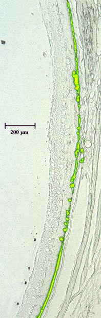
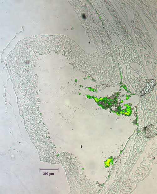
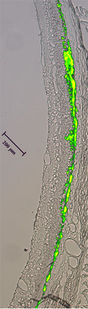
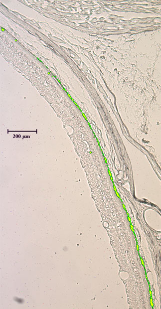
Figure 3. Sections of rat eyes after injection with fluorescent microspheres
Rat eyes were injected with 2 ml of fluorescent microspheres followed by sampling of rats two h post-injection (A), 1 day post-injection (B), 3 days post-injection (C), and 7 days post-injection (D). The rats were sacrificed by carbon dioxide asphyxiation followed by cardiac perfusion with 4% paraformaldehyde. Cryostat sections of 15 mm were prepared and viewed under fluorescence and bright field microscopy. A massive retinal detachment was visible immediately following the subretinal injection of the fluorescent microsphere suspension (A). A cluster of beads remained visible after fixation and sectioning, although most of the injected beads were lost during the processing of the sections. The retina was completely re-attached one day after the injection (B). The fluorescent microspheres spread over an area that comprised approximately 50% to 60% of the total retinal area (B). In subsequent days (C and D), the retinal area with fluorescent beads remained constant.
A:
B:
C:
D: