![]() Figure 5 of
Gilliland, Mol Vis 2001;
7:120-130.
Figure 5 of
Gilliland, Mol Vis 2001;
7:120-130.
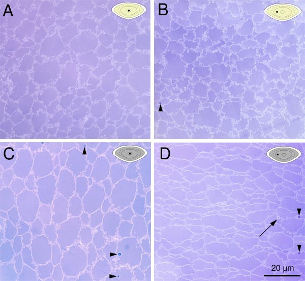
Figure 5. Light micrographs of normal and cataractous lenses
The embryonic (A) and fetal (B) nuclear regions of a normal lens appear virtually indistinguishable from the embryonic (C) and fetal (D) nuclear regions of a cataract. A multilamellar body (MLB), however, can be seen in D (arrow). MLBs occur both in normal lenses and in cataracts, but they occur with 10 times greater frequency in cataracts. Sectioning and staining artifacts are noted by arrowheads. The stain color and refractile properties visible during focusing readily distinguish the stain artifacts from MLBs. In the upper right corner of each image is a lens locator diagram (yellow for a normal lens and gray for a cataract) which contains a black dot to indicate which developmental region of the lens (see Figure 1B) is shown in the micrograph.