![]() Figure 1 of
Gilliland, Mol Vis 2001;
7:120-130.
Figure 1 of
Gilliland, Mol Vis 2001;
7:120-130.
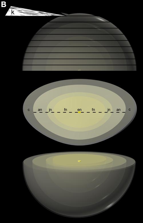
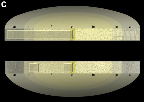
Figure 1. Diagrams of the lens processing procedure
A: Diagram of an intact human age-related nuclear cataract as would be seen in dark-field microscopy. The thin rim represents the lens capsule. The slightly yellow nucleus is shown to have radially decreasing light scattering typical of the cataractous nuclei analyzed here and pictured previously [7,9]. B: Vibratome sectioning of an intact lens into 200 mm thick slices using a thin razor blade as the knife (k). The central section through the embryonic nucleus (en) is shown in face view where all developmental regions are visible (en, fn = fetal nucleus, jn = juvenile nucleus, an = adult nucleus, and c = cortex). Sections of the nuclei from extracapsular extraction obtained in this study were bisected (dashed line) for further processing. C: Split Vibratome section from a nucleus showing the approximate cell shapes in each developmental region. Long radial mesas (upper) were used for preparing 0.5 mm thick sections for light microscopy. Cellular appearances in digitized light micrographs are superimposed on the left edge and drawings of cells are shown on the right edge. Small mesas (lower) were prepared of selected regions as a prelude to cutting 70 nm thin sections for transmission electron microscopy.