![]() Figure 10 of
Gilliland, Mol Vis 2001;
7:120-130.
Figure 10 of
Gilliland, Mol Vis 2001;
7:120-130.
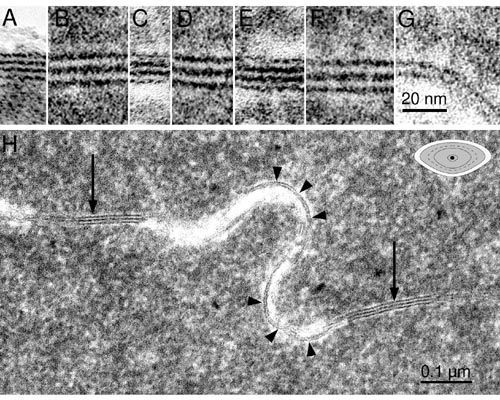
Figure 10. Comparisons of membranes by TEM
High-magnification transmission electron micrographs allow comparison of membrane thickness and staining patterns. The multilayered regions in A, C, and E are segments of multilayered membranes from MLBs, each containing three bilayers. The 5-nm distance from one lipid layer to another in these MLB membranes is probably too small to accommodate lens integral membrane proteins. The gap junctions in B, D, and F are 16 nm thick, and the single membrane in G (typical for square array junctions containing MIP/Aquaporin0) is 7 nm thick. These membranes are typical of the undulating membranes pictured in H (arrowheads). A, C, and E are taken from different segments of the MLB in Figure 9. Gap junctions in B, D, and F are taken from different gap junctions, two of which are shown in H (arrows).