![]() Figure 9 of
Gilliland, Mol Vis 2001;
7:120-130.
Figure 9 of
Gilliland, Mol Vis 2001;
7:120-130.
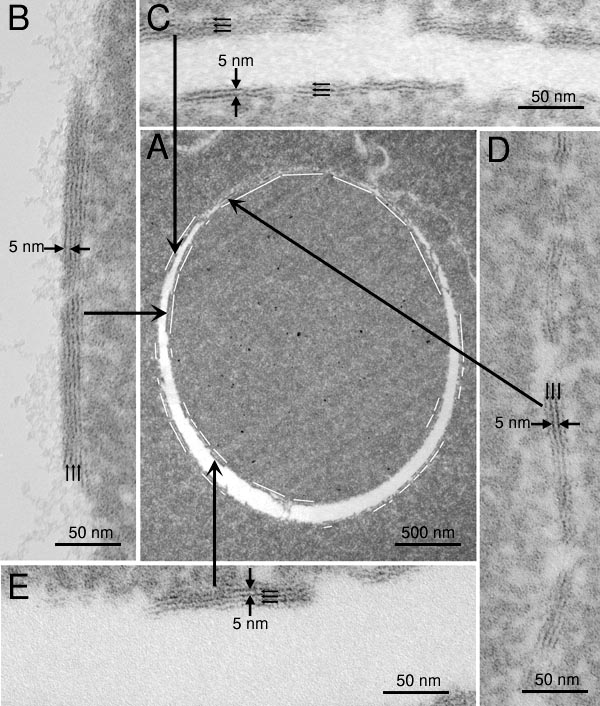
Figure 9. Electron micrographs of an MLB
Transmission electron micrographs of an MLB in a cell of the embryonic nucleus with high magnification views of membranes. Electron micrographs of one MLB (A) with insets (B, C, D, E) displaying multiple layers in different locations of the MLB. Although only a small portion of the complete layer is preserved, the geometry of the packing of the layers is distinctly different from typical membranes and junctions in the same region. The multilayered regions consistently produce a membrane thickness of 5 nm or less. The small membrane thickness is consistent with high lipid content. The white segmented line surrounding the MLB in A indicates the length and location of the visible multilayered lipid membranes within the space forming the circular border of the MLB. Short parallel arrows mark the hydrophobic interiors of adjacent lipid bilayers within multilamellar stacks.