![]() Figure 7 of
Lyubarsky, Mol Vis 2001;
7:71-78.
Figure 7 of
Lyubarsky, Mol Vis 2001;
7:71-78.
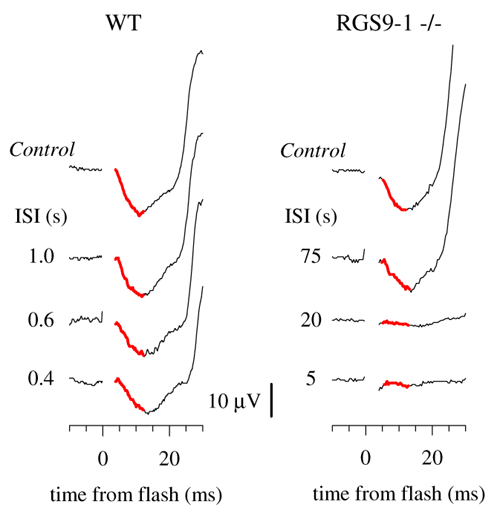
Figure 7. Recovery of the a-wave component of the ERG under cone isolation conditions for WT and RGS9-1 -/- mice
The experimental design and format of presentation were as for the results presented in Figure 6, except that the time base and amplitude scales have been expanded to reveal the initial, corneal-negative portion of the responses. In each panel, the portion of the traces identified with the suppression of cone circulating current, i.e., the cone a-wave, has been emphasized by thickening of the trace and coloring it red. The traces of the WT mouse are the same as those shown in Figure 6; those of the RGS9-1 -/- mouse were taken from a different animal from that of Figure 6, obtained in an experiment arranged to minimize the flash artifact. Each trace is the average of 10-15 records. (For clarity, a time gap of 3.5 ms containing the flash artifact has been omitted from the traces.)