![]() Figure 9 of
Gross, Mol Vis 2000;
6:51-62.
Figure 9 of
Gross, Mol Vis 2000;
6:51-62.
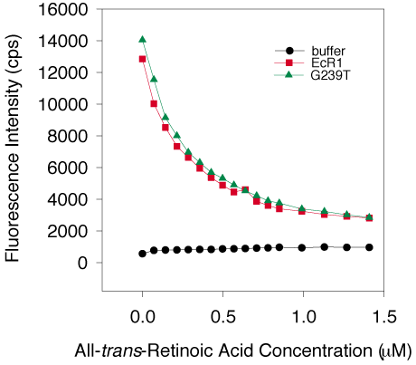
Figure 9. All-trans-Retinoic acid binding by EcR1 and G239T
Three separate G239T preparations (average represented by the green triangles) and two individual EcR1 protein preparations (average represented by the red squares), all at 1 mM, were titrated with all-trans-retinoic acid and the quenching of intrinsic protein fluorescence by this ligand was measured as described in Methods and Figure 8. Background fluorescence was examined by adding ligand to buffer without protein and is represented by the black circles. Both proteins demonstrated the same large amount of quenching (~64%) of initial fluorescence by all-trans-retinoic acid following the same curve, and saturation occurred at about 1 mM ligand, suggesting very similar specific binding of this ligand to each protein.