![]() Figure 10 of
Gross, Mol Vis 2000;
6:51-62.
Figure 10 of
Gross, Mol Vis 2000;
6:51-62.
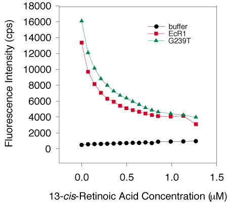
Figure 10. 13-cis-Retinoic acid binding by EcR1 and G239T
Two separate G239T preparations (average represented by the green triangles) and two individual EcR1 protein preparations (average represented by the red squares), all at 1 mM, were titrated with 13-cis-retinoic acid and the quenching of intrinsic protein fluorescence was measured as described in Methods and Figure 8. The black circles represent the background fluorescence of ligand added to buffer without protein. Both proteins demonstrated specific ligand binding as each exhibited about 68% of the initial fluorescence was quenched and saturation achieved at a concentration of ligand below 1 mM. As considered in the Discussion, the similarity in the binding curves suggest that this ligand bound equally well to each protein and that the binding site for 13-cis-retinoic acid could be identical in the two proteins.