![]() Figure 5 of
Hobby, Mol Vis 2000;
6:72-78.
Figure 5 of
Hobby, Mol Vis 2000;
6:72-78.
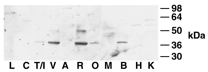
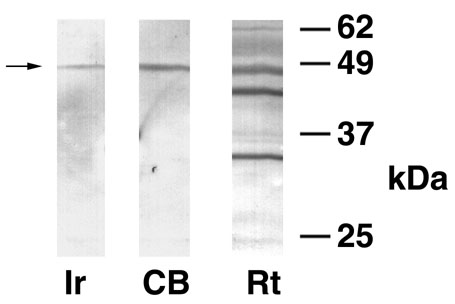
Figure 5. Western blots for human and rat tissues using a specific peptide antibody for Optc
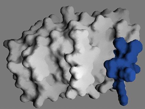
A. Location of the antigenic peptide CDPEEHKHTRRQ on a surface display of the Optc model. The residues (colored blue) form a highly accessible patch adjacent to the groove made by outer segment E'. The LRR core domain is 3.0 nm across the horizontal axis, and 2.0 nm across the vertical axis. The opticin core domain is smaller than that of the decorin model, which has 10 LRR sequence repeats [15]. B. Western blot of human eye tissues using antiserum OCGp1. Ir, iris; CB, ciliary body; Rt, retina. C. Western blot of rat tissues. L, lens; C, cornea; T/I, trabecular meshwork/iris; V, vitreous; A, aqueous; R, retina; O, optic nerve head; M, skeletal muscle; B, brain; H, heart; K, kidney.
A.
B.
C.