![]() Figure 2 of
Hobby, Mol Vis 2000;
6:72-78.
Figure 2 of
Hobby, Mol Vis 2000;
6:72-78.
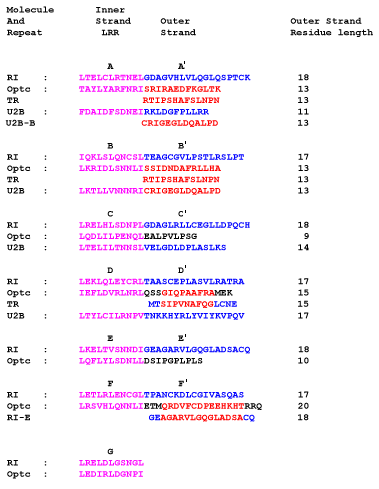
Figure 2. Sequence alignment of the seven LRR repeats of the LRR domain of Optc
The sequence alignment of the seven LRR repeats of the LRR domain of Optc is shown with structural model templates RI, TR (thyrotropin receptor), and U2A. The LxxLxLxxNxL motifs are indicated in violet and labelled A-G. Inter-LRR regions, comprising the outer segments of the molecule, are shown in blue/red/black and labelled A'-F'. Regions that have been assigned directly from one molecule to another are depicted in red as described in the text and regions that have been modeled by conformational search are in black.