![]() Figure 9 of
Kuszak, Mol Vis 1999;
5:7.
Figure 9 of
Kuszak, Mol Vis 1999;
5:7.
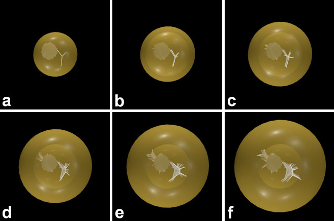
Figure 9. Scale 3D-CADS of abnormal anterior and posterior suture planes formed as a function of age during internalization of PSC
The evolution of small, triangular and rhombic suture planes produced by the overlap of the atypical suture branches in successive growth shells is shown at (A) 2 months, (B) 3 months, (C) 6 months, (D) 9 months, (E) 12 months, and (F) 15 months of age. Comparing this figure to Figure 6 and Figure 7 illustrates that the overlaying of abnormal suture patterns from successive growth shells produces abnormal suture planes. The temporal nature of suture malformation during PSC internalization is shown to greatest advantage in animated form in Figure 8.