![]() Figure 8 of
Kuszak, Mol Vis 1999;
5:7.
Figure 8 of
Kuszak, Mol Vis 1999;
5:7.
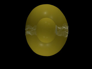
Figure 8. Scale 3D-CAD animation showing the abnormal anterior and posterior suture planes formed during PSC internalization
Over the course of one year following retinal degeneration, continuous lens growth in RCS rats results in the overlap of atypical suture patterns in successive growth shells. Thus, multiple, small, triangular and rhombic, abnormal anterior and posterior suture planes are produced and aligned directly along the visual axis.
Note that the slide bar at the bottom of the quicktime movie can be used to manually control the flow of the movie. If you do not want to or are unable to view the movie, a representative frame is included below as a still image.