![]() Figure 4 of
Thulin, Mol Vis 1999;
5:40.
Figure 4 of
Thulin, Mol Vis 1999;
5:40.
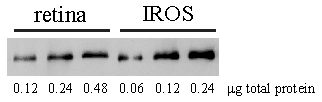
Figure 4. Determination of the concentration of rhodopsin in retinal extract
Retinal and IROS extracts prepared as described in Figure 3. Total protein loads as indicated were electrophoresed and blotted (first three lanes, retinal extract; next three lanes, IROS extract). Blot was probed with anti-rhodopsin R4 antibody. Immunoblot signal was quantitated and the concentration of Rho in retinal extract was calculated by relating to the concentration of Rho in IROS, determined by photobleaching (see text). Representative of duplicate experiments.