![]() Figure 3 of
Thulin, Mol Vis 1999;
5:40.
Figure 3 of
Thulin, Mol Vis 1999;
5:40.
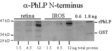
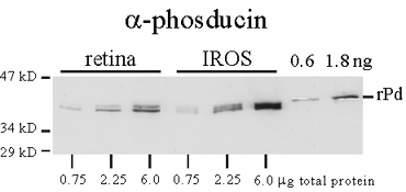
Figure 3. Phosducin and PhLP in extracts of retina and intact rod outer segments (IROS)
Bovine retina and IROS were extracted in 1% SDS, diluted to 0.3 mg/ml total protein, and amounts indicated were electrophoresed and blotted (first three lanes, retinal extract; next three lanes, IROS extract). The last two lanes contain 0.6 and 1.8 ng, respectively, of recombinant His6-Pd and His6-PhLP. Blots were probed with anti-phosducin (A) and anti-PhLP N-terminus/GST fusion protein antibodies (B). Quantitation of immunoblot signal from extract dilutions was compared with recombinant proteins to calculate the concentration of Pd and the concentration of PhLP with respect to total protein (see text).
A. Anti-phosducin antibodies.
B. Anti-PhLP N-terminus/GST fusion protein antibodies.