![]() Figure 2 of
Hageman, Mol Vis 1999;
5:28.
Figure 2 of
Hageman, Mol Vis 1999;
5:28.
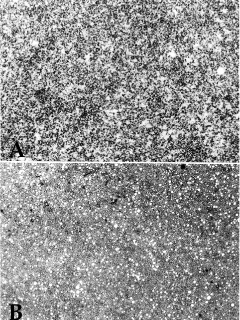
Figure 2. High magnification comparison of the first and second drusen phenotypes
High magnification transmission electron micrograph depicting the first (A; see also Figure 1) and second (B; see also Figure 3) drusen phenotypes. Whereas both classes are comprised of 20 nm granular material (A and B), the second phenotype (B) also contains small (80 nm), electron lucent, spherical elements.