![]() Figure 3 of
Hawes, Mol Vis 1999;
5:22.
Figure 3 of
Hawes, Mol Vis 1999;
5:22.
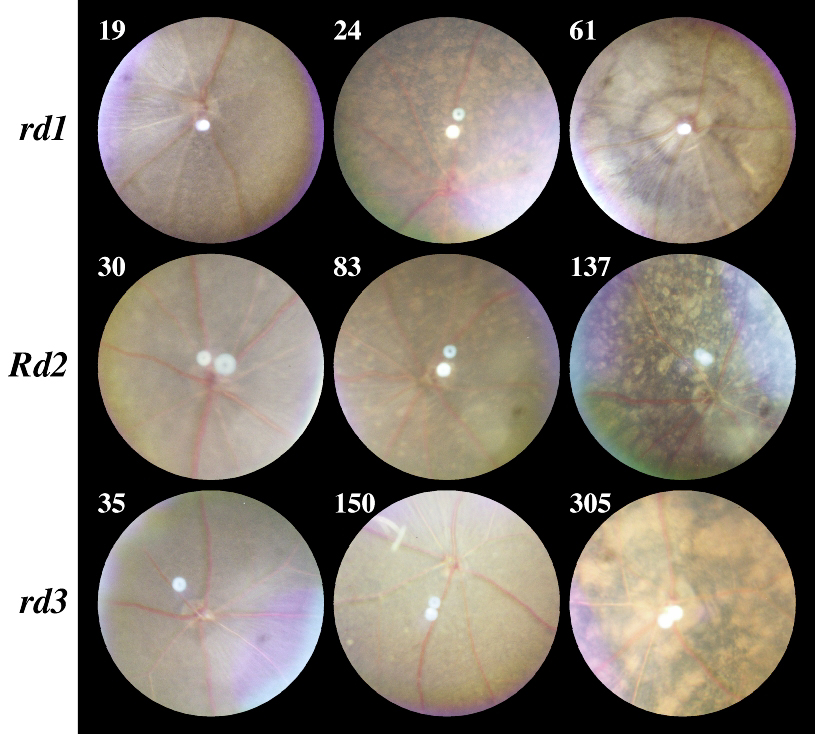
Figure 3. Progressive retinal changes
Numbers represent age in days. Top panel: retinal degeneration 1 (rd1). At 19 days the retina is similar to the normal C57BL/6J (see Figure 2), but there is narrowing of the retinal arterioles. By 24 days there is further narrowing of retinal vessels. Focal patches of RPE depigmentation are easily identified. By 61 days, all retinal vessels are narrowed and there are map-like areas of hypo- and hyperpigmentation. Middle panel: retinal degeneration 2 (Rd2). Vascular narrowing is present by 30 days of age. Development of spots of retinal pigment epithelium (RPE) depigmentation occurs by day 83. By day 137 the retina has a spotted appearance caused by areas of hypo- and hyperpigmentation of the RPE. Additional vascular narrowing is present. Bottom panel: retinal degeneration 3 (rd3). At 35 days, only vascular narrowing is seen. The degeneration has progressed by 150 days and there are small inconspicuous hypopigmented spots. In older mice (305 days) the areas of hypo- and hyperpigmentation of the RPE are prominent.
For a larger version of an individual image, click on the appropriately named text link below.
rd1, day 19 - rd1, day 24 - rd1, day 61