![]() Figure 2 of
Hawes, Mol Vis 1999;
5:22.
Figure 2 of
Hawes, Mol Vis 1999;
5:22.
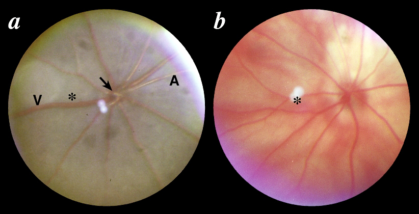
Figure 2. Normal fundi
A: C57BL/6J mouse, 8 weeks of age. The venules (V) are twice the diameter of the arterioles (A) and both demonstrate a clear blood column. The choroid and retina are pigmented. Focal faint patches of lighter pigmentation are evident. B: BALB/cByJ mouse, 12 weeks of age. Because of the absence of choroidal and retinal pigment epithelial pigmentation, the larger choroidal vessels show through the retina and the optic nerve is less well defined than in pigmented mice. Both fundi are from the right eye. The optic nerve head is indicated by an arrow and the central retina by an asterisk.
For a larger version of an individual image, click on the appropriately named text link below.
A - B