![]() Figure 2 of
Jablonski, Mol Vis 1999;
5:16.
Figure 2 of
Jablonski, Mol Vis 1999;
5:16.
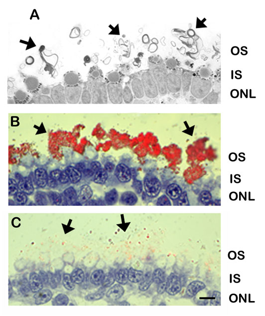
Figure 2. Morphology and immunolocalization patterns in degenerating retinas induced by RPE removal.
A. In the absence of the RPE, photoreceptor outer segments are elaborated as whorl-like structures, forming a membranous mat at the outer retinal surface (arrows). Outer segments are not localized to individual inner segments, as seen in control conditions (see Figure 1A). B. The opsin immunolabeling pattern is also indicative of this altered conformation of outer segment membranes. Individual opsin-positive outer segment profiles are not distinguishable. Rather, an irregularly shaped semi-continuous band of immunoreactivity is present (arrows). C. The pattern of rds/peripherin labeling was very irregular and much of it is out of the plane of focus, reflecting the disordered array of outer segments (arrows). Bar = 10 µm.